X-Ray
X-ray, or radiography, is the oldest and most common form of medical imaging. An X-ray machine produces a controlled beam of radiation, which is used to create an image of the inside of your body. This beam is directed at the area being examined.

Color Doppler
This technique estimates the average velocity of flow within a vessel by color coding the information. The direction of blood flow is assigned the color red or blue, indicating flow toward or away from the ultrasound transducer.
UltraSound
Medical ultrasound (also known as diagnostic sonography or ultrasonography) is a diagnostic imaging technique based on the application of ultrasound. It is used to see internal body structures such as tendons, muscles, joints, blood vessels, and internal organs.

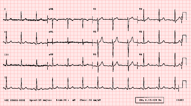
ECG
A recording of the electrical activity of the heart. Abbreviated ECG and EKG. ... Electrodes are placed on the skin of the chest and connected in a specific order to a machine that, when turned on, measures electrical activity all over the heart.